Intraoral Camera
Grants Pass, OR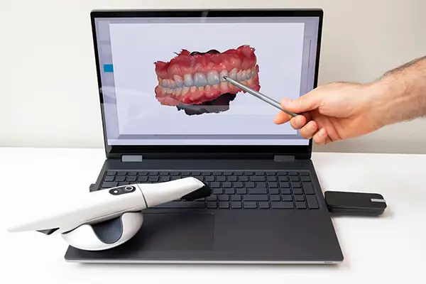 Patients often arrive at a dental appointment with at least one question in mind: how does this compare to what dentistry used to look like? Many people remember sitting through x-rays as the primary way a dentist checked for cavities or other concerns. Today, intraoral cameras work alongside x-rays to give a more complete picture of what is happening in the mouth. Understanding how these two tools differ, and how they complement each other, helps explain why The Center for Esthetic Dentistry relies on both during a thorough exam. Patients often arrive at a dental appointment with at least one question in mind: how does this compare to what dentistry used to look like? Many people remember sitting through x-rays as the primary way a dentist checked for cavities or other concerns. Today, intraoral cameras work alongside x-rays to give a more complete picture of what is happening in the mouth. Understanding how these two tools differ, and how they complement each other, helps explain why The Center for Esthetic Dentistry relies on both during a thorough exam.What X-Rays ShowDental x-rays remain an essential diagnostic tool because they can see what the eye and the camera cannot. X-rays penetrate through tooth enamel and bone, revealing decay between teeth, infections beneath the gumline, bone loss, and the position of teeth that have not yet erupted. This internal view is something no camera, no matter how advanced, can replicate, which is why x-rays continue to play a central role in a comprehensive dental exam at our practice. What Intraoral Cameras ShowIntraoral cameras, by contrast, capture what is visible on the surface of the teeth and gums, but in far greater detail than the naked eye can manage. Where an x-ray shows shadows and densities, the camera shows full color, real texture, and surface-level detail such as cracks, stains, early erosion, and inflamed or receding gum tissue. The two technologies are not competing with each other. They are answering different questions about the same mouth. Comfort And ConvenienceOne practical difference patients notice is comfort. X-rays require a sensor or film to be positioned inside the mouth, sometimes held in place with a bite tab, and the process can feel slightly awkward for a few seconds at a time. Intraoral cameras, on the other hand, involve nothing more than a small wand gliding gently along the teeth, with no biting, no holding still in an uncomfortable position, and no exposure to radiation. For patients who find x-rays mildly unpleasant, the camera portion of the exam tends to feel far more relaxed. Real Time Viewing Versus Developed ImagesAnother meaningful difference is how quickly each image becomes useful. Digital x-rays do appear on screen fairly quickly, but they still require some degree of interpretation, since the resulting image is a black and white representation of internal structures. Intraoral camera footage, by comparison, streams live to the monitor as the wand moves, so your dentist can narrate findings to you in real time, pointing at exactly what is being discussed on a full color image that needs no special training to interpret. X-rays vs IntraOral Cameras:
Why Both Tools Matter TogetherA thorough dental exam benefits from combining both perspectives. If an x-ray reveals a shadow suggesting decay between two teeth, an intraoral camera image of the surrounding area can help confirm visible signs and give a clearer sense of how far a problem may have progressed. Likewise, if the camera reveals a crack or area of concern on the surface, an x-ray can help determine whether that issue extends below the gumline or into the supporting bone. Used together, the two technologies reduce the chances that something gets missed. How Often Each Is UsedBecause x-rays involve a small amount of radiation, they are typically taken on a recommended schedule rather than at every single visit, often once a year for healthy adult patients, with more frequent imaging when a specific concern needs monitoring. Intraoral camera images, since they involve no radiation at all, can be captured as often as needed without any added risk, which makes them a natural tool for closer, more frequent monitoring between x-ray appointments. A More Complete Picture Of Your Oral HealthWhen patients understand the distinct roles these two technologies play, the reasoning behind a thorough exam becomes much clearer. X-rays give us the internal view that protects against problems hiding beneath the surface, while intraoral cameras give us, and you, a detailed external view that supports day to day monitoring and clear communication. Together, they allow our dental team to build a far more complete picture of your oral health than either tool could provide on its own. Helping Patients Understand Their Own ExamOne often overlooked difference between these two tools is how each one involves the patient in the exam itself. An x-ray, once captured, is typically reviewed by the dentist and explained afterward, since interpreting the grayscale image takes training most patients do not have. An intraoral camera image, on the other hand, requires no special interpretation. A crack looks like a crack. Inflamed gum tissue looks visibly different from healthy tissue. This makes the camera a particularly useful teaching tool during the exam itself, since patients can follow along and ask questions about exactly what they are seeing in the moment, rather than waiting for a summary afterward. Supporting Long Term MonitoringMany dental concerns develop gradually, which means tracking change over time is often more useful than any single image on its own. X-rays taken at routine intervals allow your dentist to compare bone levels or watch a small area of decay over successive visits. Intraoral camera images serve a similar purpose for surface conditions, allowing direct visual comparison of how a stained area, a worn edge, or a patch of gum recession has changed from one appointment to the next. Having both forms of documentation creates a more reliable long-term record than relying on memory or written notes alone. Two Tools, One GoalNeither tool exists to replace the other, and a thorough exam rarely relies on just one. By combining the internal clarity of x-rays with the surface detail and real time communication of intraoral cameras, our team approaches each patient's care with a more complete, more confident understanding of what is actually happening in their mouth. At The Center for Esthetic Dentistry, we use both intraoral cameras and digital x-rays to give every patient a thorough, well rounded exam. If you have questions about either technology or would like to schedule a visit, call us at 541-476-8788. We are happy to walk you through exactly what each tool shows us about your smile. |
 Hours Mon Tues Weds Thur Fri Sat - Sun 8am to 5pm 8am to 5pm 8am to 5pm 8am to 5pm 8am to 5pm Closed |
