Digital X-Ray
Grants Pass, OR
Patients preparing for a dental appointment with a dental cleaning and dental exam often have questions about X-rays that go beyond what the images show. How often are they actually needed? Is the radiation something to be concerned about? What determines whether a patient needs a full series or just a couple of images? At The Center for Esthetic Dentistry we believe these are practical questions that deserve straightforward answers. Understanding the factors that guide X-ray frequency, the safety profile of digital dental imaging, and what the process involves from the patient's perspective makes it easier to approach dental care with accurate expectations. Why X-Ray Frequency Is Not the Same for Every PatientThere is no universal answer to how often dental X-rays should be taken because the appropriate frequency depends on the individual patient's oral health status, history, and risk factors. Professional guidelines from organizations like the American Dental Association recommend that X-ray frequency be determined by the clinical judgment of the treating dentist based on each patient's specific circumstances, rather than following a fixed schedule that applies to everyone. A patient who has had no cavities in years, maintains excellent home care, and has stable gum health may need bitewing X-rays only every two to three years. A patient who is prone to decay, is being treated for gum disease, or has a history of complex restorative work may need more frequent imaging to monitor changes in a timely way. New patients typically receive a comprehensive set of X-rays so the dentist has a complete baseline picture of the mouth before recommending a routine imaging schedule. Types of X-Rays and When Each Is UsedThe type of X-ray taken at any given appointment is determined by what needs to be evaluated, not by routine.
Understanding Radiation Exposure in PerspectiveRadiation is a word that can create anxiety out of proportion to the actual risk involved in dental X-rays. Context helps. Radiation exists naturally in the environment in the form of cosmic rays, soil, building materials, and food. Every person receives a baseline level of radiation exposure simply by living on Earth, a figure called background radiation. The radiation from a full set of digital dental bitewing X-rays is equivalent to, or less than, what a person naturally absorbs from the environment in a single day. Digital X-rays have further reduced exposure compared to traditional film X-rays, which were already low-risk. The sensors used in digital imaging require significantly less radiation to produce a high-quality image than film required, and the speed of digital capture means the exposure duration is minimized. Protective lead aprons placed over the patient's chest and lap during imaging provide additional shielding, and thyroid collars are available for patients who prefer them. X-Rays and PregnancyPregnant patients may wonder whether dental X-rays are safe during pregnancy. The general guidance from dental and obstetric organizations is that routine dental care, including X-rays when clinically necessary, is safe during pregnancy. The radiation exposure from dental X-rays is localized to the head and neck area, and with a lead apron in place, exposure to the abdominal area is negligible. Delaying necessary dental treatment during pregnancy due to concerns about X-rays can sometimes lead to greater problems. Untreated dental infections and gum disease have been associated with adverse pregnancy outcomes, making timely diagnosis and treatment genuinely important. Patients who are pregnant should always inform their dental team so that precautions can be taken and any X-rays are used only when clinically warranted. What to Expect During the Imaging ProcessDigital X-rays are a brief and straightforward part of the dental appointment. The dental assistant or hygienist will position a small sensor inside the mouth, typically in the area of the teeth being imaged. The patient is asked to hold still while the X-ray is taken, which takes less than a second. The image appears on the chairside monitor almost immediately, and the sensor is repositioned as needed for each additional view. Patients with a strong gag reflex sometimes find intraoral sensors uncomfortable, particularly when positioned toward the back of the mouth. Informing the dental team ahead of time allows them to use strategies that minimize discomfort, including sensor size selection, distraction techniques, or adjusting positioning. Phosphor plate sensors, which are thinner and more flexible than rigid digital sensors, may be used for patients who have difficulty tolerating a firmer device. Reviewing Images With Your Dental TeamOne of the meaningful differences digital X-rays make in the patient experience is the ability to review images together in real time. Once captured and displayed on the monitor, the dentist or hygienist can walk the patient through each image, pointing out areas of concern, explaining what healthy bone and tooth structure look like, and comparing current images to those from previous visits. This immediate review encourages patients to ask questions and participate actively in understanding their oral health. Seeing the reason behind a recommendation, such as a dark spot between two teeth indicating early decay, makes it easier to understand why treatment is being suggested and what happens if it is deferred. Keeping Records Accurate Over TimeDigital X-rays are stored in the patient's electronic health record and remain accessible across every subsequent visit. When a patient transfers from another practice, requesting the digital files is simple and ensures continuity of care without a gap in imaging history. Over the course of years, those stored images provide a meaningful longitudinal record of how a patient's oral health has evolved, which supports more accurate diagnosis and more informed treatment planning. Patients who have had X-rays taken at previous offices are encouraged to request copies of those images when establishing care at a new practice. The more complete the imaging history, the better the baseline from which future changes can be measured. We're Here to Answer Your Questions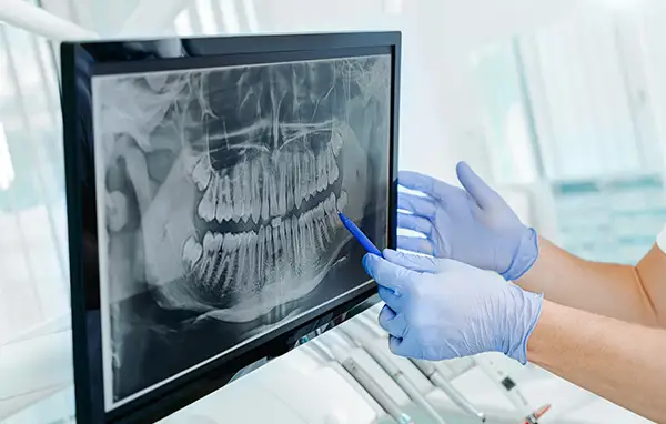
X-rays are a routine and important part of the care we provide at The Center for Esthetic Dentistry, and we welcome any questions you have about the process, the technology, or how often imaging is appropriate for your specific situation. Call our office at 541-476-8788 to schedule your appointment or to speak with a member of our team. |
 Hours Mon Tues Weds Thur Fri Sat - Sun 8am to 5pm 8am to 5pm 8am to 5pm 8am to 5pm 8am to 5pm Closed |
